After a crash, a lot of people expect the hard part to be over once the emergency room says the scan looks normal. Then the actual confusion starts. You forget words mid-sentence. Lights bother you. Your balance feels off in the grocery store. Reading makes your eyes ache. Family members may mean well and say, “At least the MRI was clear,” but that can leave you feeling more alone, not less.
That disconnect is common with traumatic brain injuries and other crash-related nerve injuries. A CT or MRI looks for structure. Your symptoms may come from a problem in function, meaning the way signals move through the nervous system. That's where evoked potential tests can matter. They don't just take a picture. They measure whether messages from your eyes, ears, or body are getting to your brain at the right speed and in the right way.
When You Feel Injured but Scans Look Normal
The most frustrating injuries are often the ones nobody can see.
You may have walked away from the crash, gone home, and assumed the dizziness would pass in a few days. Instead, weeks later, you're still struggling with screen time, headaches, sound sensitivity, blurred vision, or that unsettling “foggy” feeling that makes simple tasks feel harder than they should.

Why normal imaging can feel misleading
A normal scan doesn't mean you're imagining your symptoms. It often means the scan didn't capture the kind of problem that's affecting you.
Think about the difference between a photograph of a car and a road test. A photograph can show dents, broken glass, and bent metal. It can't tell you whether the steering pulls left, the brakes hesitate, or the engine misfires under pressure. Brain imaging can face a similar limit. It may show anatomy well but miss subtle disruption in how nerve pathways are working.
That's why doctors sometimes add tests that look at function from a different angle. If you're trying to understand how clinicians use technology to measure movement and recovery more broadly, this physical therapist technology guide gives a useful overview of how tools can capture problems that aren't obvious to the naked eye.
Why this matters after a crash
After head trauma, many people develop symptoms that sound subjective to outsiders. “I feel off.” “My vision isn't right.” “I get dizzy when I turn my head.” Those experiences are real, but they can be hard to prove unless testing picks up something objective.
Evoked potential tests can help fill that gap. They measure the nervous system's electrical response to a controlled stimulus, such as a visual pattern, a sound, or a mild sensory input. Instead of asking only what the brain looks like, the test asks whether signals are traveling normally.
What many patients need to hear: A normal scan and a real injury can exist at the same time.
In some cases, doctors may combine different advanced tools to get a fuller picture. Another example is diffusion tensor imaging of the brain, which looks at white matter pathways differently than standard imaging. The larger point is simple. If your symptoms continue, the evaluation shouldn't stop just because the first scans looked clean.
How Evoked Potential Tests Measure Nerve Function
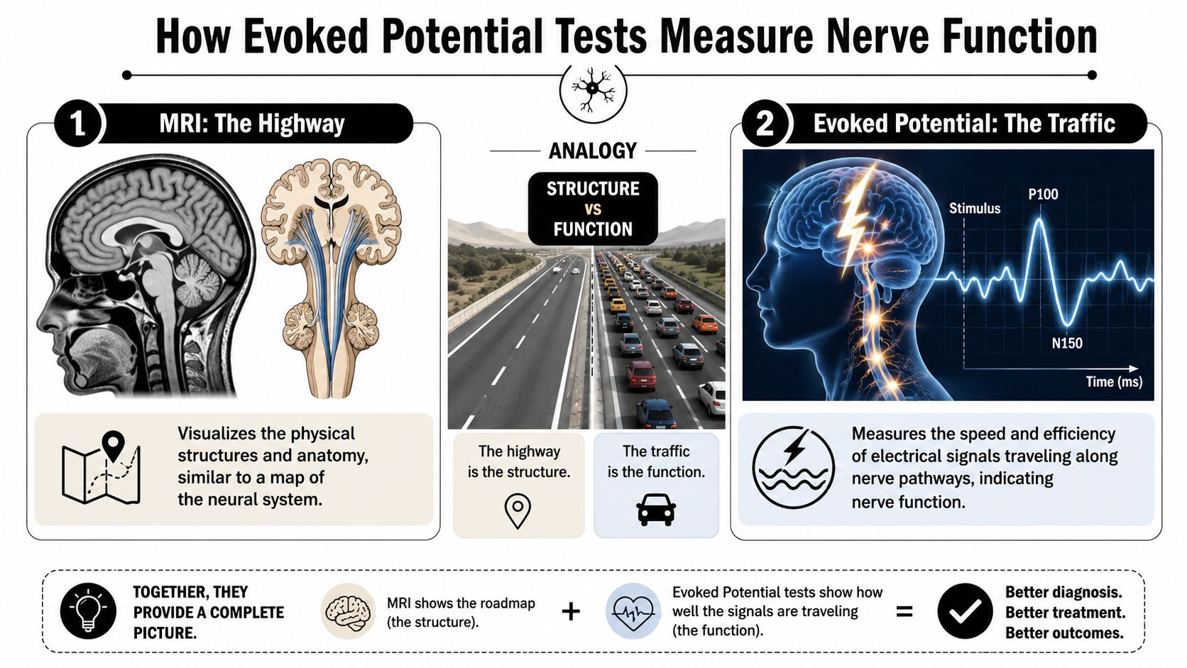
An evoked potential test works a bit like checking signal speed on a network.
A regular scan shows the wiring. An evoked potential test checks how quickly a message moves through that wiring. If the message is delayed, distorted, or weak, that can point to dysfunction along the pathway even when the structure itself doesn't show an obvious problem.
The basic idea
During the test, a clinician gives your nervous system a specific stimulus. Then electrodes placed on your scalp and sometimes other parts of your body record the response. The key question is whether the response arrives when it should.
Here's the plain-language version:
- Visual stimulus: You look at a changing pattern or light, and the test tracks how the visual pathway responds.
- Auditory stimulus: You hear clicks or tones, and the test tracks signal travel through the hearing and brainstem pathways.
- Sensory stimulus: You receive a mild physical stimulus, and the test tracks how signals move from the body toward the brain.
If the pathway is healthy, the response follows an expected pattern. If the pathway has been disrupted, the response may arrive later than expected or look different in size and shape.
Why doctors trust this method
These tests aren't experimental. According to Cleveland Clinic's evoked potential test overview, evoked potential tests have been in clinical use for over a century, and the modern diagnostic era took off in the 1970s when EEG-based averaging made it possible to detect tiny brain signals hidden in background noise. That same source explains that today's tests are standardized to measure signal travel time from the eyes, ears, or limbs to the brain.
That history matters when you're already overwhelmed. It means your doctor isn't reaching for an unusual tool. They're using an established method for testing how nerve pathways function.
A scan can answer “Do we see damage?” An evoked potential test can answer “Is the pathway working properly?”
Why this can be useful in brain injury workups
Traumatic brain injury symptoms often involve slowed processing. You may notice that your eyes need longer to focus, your balance reactions feel delayed, or sounds overwhelm you in a busy room. Those experiences all involve communication inside the nervous system.
An evoked potential test doesn't measure every possible symptom after a crash. But it can reveal whether certain sensory pathways are conducting signals normally. If you're comparing this test with other tools doctors use in head injury cases, this overview of neurological tests for brain damage helps place it in context.
The Three Main Types of Evoked Potential Tests
Doctors usually choose the test based on your symptoms, not just the fact that you were in a crash. That matters because each test checks a different pathway.
Visual evoked potential
A visual evoked potential, often called VEP or VER, looks at how signals travel from the eyes through the optic pathways to the brain.
If you've been dealing with blurred vision, trouble reading, eye strain, or a feeling that your brain can't keep up with what your eyes are seeing, this is often the type people hear about first. The test usually involves watching a screen while electrodes record the brain's response.
For a crash victim, this can help when the complaint isn't “I can't see” in the usual sense. It's often more subtle than that. You may still read the eye chart, but screens trigger headaches, tracking moving objects feels harder, or visual tasks leave you exhausted.
Brainstem auditory evoked response
A brainstem auditory evoked response, also called BAER or BAEP, focuses on hearing-related pathways and the brainstem.
This can be relevant if you have ringing in the ears, unusual sound sensitivity, dizziness that seems tied to motion or auditory overload, or a sense that your hearing is technically present but not processing correctly. During the test, you usually listen to sounds through earphones while electrodes record how the auditory system responds.
This type often confuses patients because they assume it's only for obvious hearing loss. It isn't. The test is about how sound signals move through the pathway, especially through the brainstem, which can matter after head trauma.
Somatosensory evoked response
A somatosensory evoked response, called SSEP or SSER, tracks signals traveling from the body toward the brain.
Doctors may order this if you have numbness, tingling, altered sensation, weakness, or concern about spinal cord pathway injury after a crash. The stimulus is typically a mild electrical input to a peripheral nerve, and the recording shows how that signal travels centrally.
If your symptoms involve the neck, arms, hands, legs, or a patchy sense that one side of your body feels “different,” this test can help identify whether the sensory pathway is conducting normally.
Comparing the three main evoked potential tests
| Test Type | Stimulus Used | What It Tests | Symptoms Investigated |
|---|---|---|---|
| Visual Evoked Potential (VEP/VER) | Visual pattern or light | Visual pathway from eyes to brain | Blurred vision, visual strain, reading difficulty, post-traumatic visual symptoms |
| Brainstem Auditory Evoked Response (BAER/BAEP) | Sound such as clicks or tones | Auditory pathway and brainstem function | Dizziness, tinnitus, sound sensitivity, auditory processing concerns |
| Somatosensory Evoked Response (SSEP/SSER) | Mild sensory or electrical stimulation to a nerve | Sensory pathways from limbs/body to brain | Numbness, tingling, weakness, sensory changes, suspected spinal pathway problems |
One test doesn't answer every question
A lot of confusion comes from assuming “an evoked potential test” is one single exam. It's really a family of tests. The one your doctor orders should match the symptoms you're having.
If your symptoms are visual, auditory, balance-related, or sensory, the pathway tested should fit that complaint. That matching process is part of what makes the result meaningful.
Why Your Doctor Ordered This Test After a Crash
Crash injuries don't always leave behind obvious structural damage on standard imaging. But they can still disrupt the brain and spinal pathways that carry information from one place to another.
Functional injury after violent motion
In a car or truck collision, the brain and body can be exposed to sudden acceleration, deceleration, rotation, and impact. Even if you never lost consciousness, those forces can affect delicate nerve fibers and the way electrical signals travel along them.
That's one reason a person can look “fine” on paper but feel very different in daily life. They may struggle with concentration, balance, vision, hearing-related sensitivity, or limb sensations that started only after the collision. The issue may be less about a large visible lesion and more about disrupted signaling.
Why a doctor picks this test instead of using it for everyone
Evoked potential tests are highly specific. They aren't broad routine screenings for every patient with every complaint. According to UMass Memorial Health's overview of evoked potential testing, their value is tied closely to the clinical question, and use is growing in complex settings such as monitoring spinal cord function during surgery and evaluating brain activity in coma patients to help predict recovery.
That selectivity is helpful in a crash case. It means your doctor usually isn't ordering the test casually. They're ordering it because your symptoms suggest a particular pathway may have been affected.
How that connects to traumatic brain injury
With traumatic brain injury, people often hear, “Your MRI didn't show anything serious.” That can sound reassuring, but it doesn't always answer why you still can't tolerate motion in a busy store or why reading makes you nauseated.
Evoked potential tests can provide objective physiologic evidence that a pathway isn't conducting normally. That doesn't replace the rest of your medical evaluation. It adds a layer of evidence that can support what you've been feeling all along.
A few examples of when that logic fits after a crash:
- Visual complaints after head impact: A visual pathway test may help if your vision feels altered despite a normal eye exam.
- Dizziness with sound sensitivity or ringing: An auditory brainstem test may help explore whether the issue involves more than the ear itself.
- Numbness or altered body sensation: A somatosensory study may help identify a pathway problem involving the spine or sensory tracts.
The point of this test isn't to “prove you're injured enough.” It's to measure whether a nerve pathway is functioning the way it should.
What to Expect During Your Testing Appointment
Individuals often feel less anxious once they understand the appointment is quiet, noninvasive, and structured.
According to the clinical overview from Cleveland Clinic cited earlier, these studies are commonly done as outpatient procedures lasting about 60 to 90 minutes, with interpretation by neurologists or neurophysiologists. That gives you a rough sense of the day without making the visit feel mysterious.
What happens first
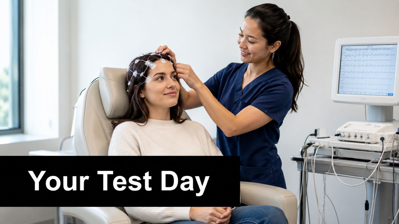
When you arrive, a technician will usually explain which pathway is being tested and place small electrodes on your scalp and sometimes other areas of your body. These electrodes record electrical activity. They don't shock your brain.
The paste or gel can feel cool or sticky, but the setup itself is generally painless. The room may be dim or quiet so the recording is cleaner and distractions are reduced.
What the test feels like
The sensation depends on the type of evoked potential test:
- For visual testing: You may watch a screen and focus on a pattern.
- For auditory testing: You may wear earphones and listen to repeated sounds.
- For somatosensory testing: You may feel mild stimulation on an arm or leg while the system records the response.
The hardest part for many patients isn't pain. It's staying still and attentive enough for the recording to be useful. If you have a headache, nausea, light sensitivity, or fatigue after your crash, tell the staff. They can often help you pace the test.
What happens after
Once the electrodes are removed, you can usually leave right away. There isn't typically a recovery period just from the test itself.
Practical rule: Ask before you leave when and how results will be explained. A technical report by itself can be hard to interpret without a clinician walking you through it.
Understanding Your Test Results and Their Limits
The report may look intimidating at first. It can include wave labels, timing values, and technical language that doesn't sound anything like the symptoms you've been living with.
The two core ideas are latency and amplitude.
Latency means timing
Latency is the time it takes for the signal to travel from the stimulus point to the part of the nervous system being measured. If the response arrives later than expected, that can suggest slowed conduction somewhere along the pathway.
The University of Iowa Health Care explains that evoked potential tests measure the timing, or latency, and the size, or amplitude, of a nerve response, and that a delay as small as 10 milliseconds may be significant for nerve pathway damage in the right clinical setting, as noted in its evoked potential test guidance.
That matters because many crash injuries are subtle in daily life. You may function, but more slowly. A delayed response can provide objective support for that kind of complaint.
Amplitude means signal size
Amplitude refers to the size of the recorded response. In plain language, it reflects how strong the measured signal appears.
The interpretation of results gets more nuanced. A weak response doesn't always mean a severe injury, and a stronger response doesn't always mean everything is normal. Clinicians look at patterns, symmetry, timing, and consistency together.
A research paper available through NIH's PubMed Central notes that motor evoked potentials show substantial variability within the same person and between different people, which is why researchers developed dedicated statistical models for repeated measurements rather than treating the signal as a simple yes-or-no event. That variability is discussed in this PMC paper on motor evoked potential modeling.
Why one abnormal wave isn't the whole story
Doctors don't usually base interpretation on a single blip. They evaluate repeated responses and look for a meaningful pattern.
That's important if you're reading your own report and trying to guess what every line means. Medical records often use compressed formatting and shorthand. If you want a plain-language primer on how clinicians organize findings, this guide to understanding medical report formats can make the layout easier to follow.
An evoked potential test can support a diagnosis. It doesn't replace the bigger clinical picture, which includes your symptoms, exam, and other testing.
A normal result doesn't end the conversation
This is one of the hardest truths for patients. A normal result doesn't automatically rule out injury.
Evoked potential tests only measure the pathways they are designed to test. If your symptoms come from a different pathway, a more subtle issue, or a problem the test isn't built to detect, the result may be normal even though you still have a real post-crash condition. That's why specialists interpret these studies in context instead of treating them like a final verdict.
Using EP Tests to Strengthen Your Recovery and Claim
When symptoms are invisible, people often feel like they have to defend their own reality. That can happen in the exam room, at work, and during an insurance claim.
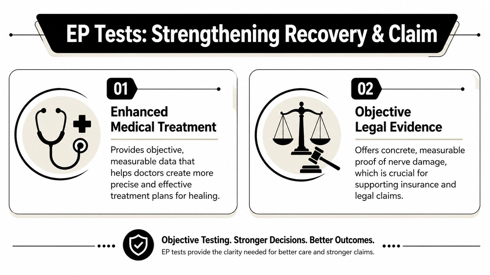
Evoked potential tests can help because they turn part of that story into objective medical data. If a visual pathway is delayed, that gives a treating doctor something concrete to work with. If a sensory pathway shows abnormal conduction, that can guide referrals, rehabilitation planning, and follow-up testing.
Why the medical side matters
Recovery gets easier to target when the problem is described clearly. Instead of a chart saying only “patient reports dizziness,” the record may show measurable dysfunction in a pathway tied to balance, vision, hearing, or sensory processing.
That can support the next steps your doctor recommends, whether that means further neurologic evaluation, vision therapy, vestibular care, or other rehabilitation.
Why the legal side matters
Claims often rise or fall on documentation. Subjective symptoms matter, but insurers tend to challenge complaints they can't see on a scan. An evoked potential test may help connect what you feel to a measurable physiologic finding.
That doesn't prove every part of a claim by itself. It does strengthen the record. And when pain, cognitive strain, dizziness, or sensory disruption affect your daily life, that evidence can be part of showing the human impact discussed in guides about how to prove pain and suffering.
A good claim file doesn't just say you hurt. It shows how your body and brain changed after the crash.
If you're dealing with post-crash symptoms that don't match what the first scans showed, Nares Law Group LLC understands how complex brain injury evidence can be. The firm helps injured people connect medical findings, ongoing symptoms, and legal proof so they can pursue the care and accountability they deserve.