When a standard MRI or CT scan comes back “normal” after a serious accident, it’s beyond frustrating. You’re living with real, debilitating symptoms, yet the tests show nothing wrong. This is where Diffusion Tensor Imaging (DTI) of the brain becomes a critical tool. It can see what other scans miss, providing objective proof of microscopic brain damage that validates your experience and can make or break your legal case.
Seeing Invisible Brain Injuries After an Accident
After a car crash or a bad fall, doctors will usually order a CT scan or a standard MRI. They’re looking for the big things—skull fractures, brain bleeds, or major swelling. When those tests are clear, insurance companies often jump to the conclusion that your headaches, memory loss, or mood swings aren’t related to the accident. For too many survivors of traumatic brain injury (TBI), this is where the real fight begins.
The problem is, conventional imaging was never designed to see damage at a microscopic level. It can’t detect the widespread, tiny tears in nerve fibers, an injury known as diffuse axonal injury (DAI). This is one of the most common outcomes of the violent forces your brain endures in an accident.
Uncovering the Hidden Damage
This is exactly why diffusion tensor imaging of the brain is so important.
Think of your brain’s white matter as a massive, intricate network of fiber-optic cables that allows different parts of your brain to communicate. A standard MRI might show if a whole bundle of cables has been cut, but it can’t tell you if the individual fibers inside are frayed, stretched, or damaged.
DTI, however, is a more sophisticated scan. It works by tracking the movement of water molecules throughout the brain’s white matter pathways.
- In a healthy brain, water moves in a predictable, organized direction along those nerve fibers.
- When those fibers are damaged, the structure breaks down. Water can no longer flow in an orderly way and instead diffuses more randomly, like water seeping out of a leaky, damaged pipe.
DTI technology measures this disruption. It creates a detailed map that quite literally shows the broken pathways inside the brain—the very damage that is causing your symptoms. To better understand how the legal system views these injuries, see our article on undetected brain injuries.
Why This Matters for Your Injury Claim
For personal injury and wrongful-death claims, this kind of objective data is a game-changer. The clinical power of DTI is especially clear in TBI assessments. While research shows that healthy aging can cause minor changes in white matter, trauma from an accident causes severe and accelerated damage. After a crash, victims with DAI can show a 20-30% drop in white matter integrity in critical areas like the corpus callosum—a drop that directly correlates with how severe their outcome will be. You can explore the full research on white matter integrity and trauma for more detail.
By visualizing what was once invisible, DTI provides clear, undeniable proof linking the accident to a physical brain injury. It turns your subjective symptoms into objective evidence. This is absolutely essential for holding the negligent party accountable and securing the compensation you need for your long-term care.
How DTI Makes Invisible Brain Damage Visible
To really get what diffusion tensor imaging does, you have to start with a simple fact: your brain is about 75% water. DTI is a highly advanced form of MRI that works by tracking how all those water molecules move—or "diffuse"—through the brain's complex network.
That movement tells a powerful story about the health of your brain's white matter, which acts as its communication wiring.
In a healthy brain, water doesn't just slosh around. It flows in a very specific, organized way along the tightly packed bundles of nerve fibers, known as axons. Think of it like a perfectly contained river flowing smoothly down a well-defined channel. The flow is directional and predictable.
This orderly, one-way flow is called anisotropy, and it's a key sign of healthy, intact brain wiring.
Spotting the Disruption with Key DTI Metrics
When the violent forces of an accident impact the head, those delicate nerve fibers can be stretched, twisted, or torn apart. This is a diffuse axonal injury, and it completely disrupts the brain's organized structure. The once-orderly pathways are now broken.
Suddenly, water molecules are no longer flowing down a clear channel. Instead, they spill out and move randomly in all directions, like water spraying from a burst pipe. The flow becomes chaotic and disorganized.
DTI technology is designed to see this exact change. To pinpoint the damage, neuroradiologists look at two primary measurements:
- Fractional Anisotropy (FA)
- Mean Diffusivity (MD)
Understanding these two numbers is the key to seeing what a DTI scan is truly showing about your injury.
Fractional Anisotropy (FA): The Directional Score
Fractional Anisotropy (FA) is the most critical number that comes out of a DTI scan. It’s a score from 0 to 1 that measures how directional the water movement is.
A high FA value, closer to 1, is good news. It means water is flowing in a single, dominant direction. This tells us the white matter tracts are dense, healthy, and working as they should—like our smooth-flowing river.
A low FA value, closer to 0, is a major red flag. It shows that water is diffusing randomly instead of in a straight line. This is strong evidence that the underlying white matter has been damaged or destroyed, breaking down the pathways that once guided the water’s flow.
In a personal injury case, a low FA value in a specific part of the brain is objective, measurable proof of axonal injury. It makes the microscopic damage that standard MRIs almost always miss completely visible.
The flowchart below shows how DTI can uncover this hidden damage when other scans come back "normal."
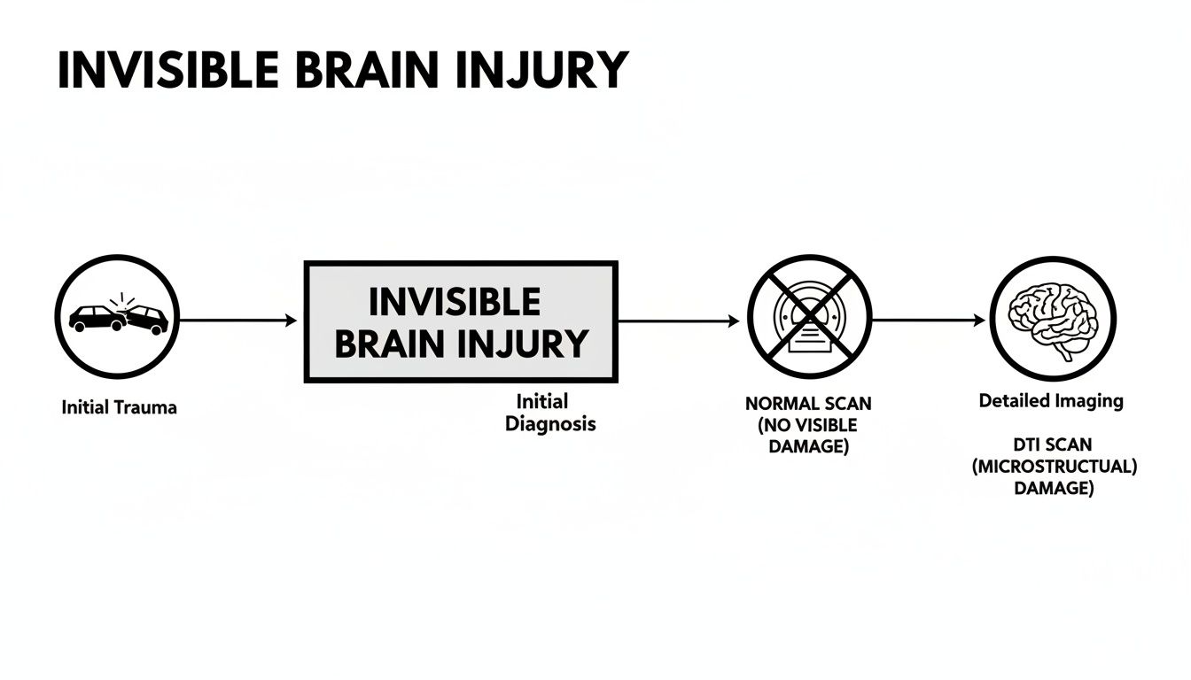
It maps out the diagnostic journey many of our clients take, where a conventional MRI shows nothing wrong, but a DTI scan finally reveals the true source of their symptoms.
Mean Diffusivity (MD): The Water Flow Volume
While FA tells us about the direction of water flow, Mean Diffusivity (MD) tells us about the total amount of water movement in an area. It measures the overall magnitude of diffusion, no matter the direction.
After a brain injury, damaged cells break down and can cause swelling (edema). This process creates more empty space in the tissue, giving water molecules more room to move around.
An abnormally high MD value often points to this kind of cellular damage and swelling. When a radiologist sees high MD in the same area as low FA, it provides a powerful, complementary picture of white matter injury. The high MD reinforces what the low FA is telling us: the tissue’s integrity has been compromised.
If you want to learn more about the different types of imaging, our guide on the differences between various brain MRI scans is a great resource.
Together, these metrics transform an injury that was once invisible into clear, data-driven proof.
What DTI Scans Reveal About Your Brain Injury
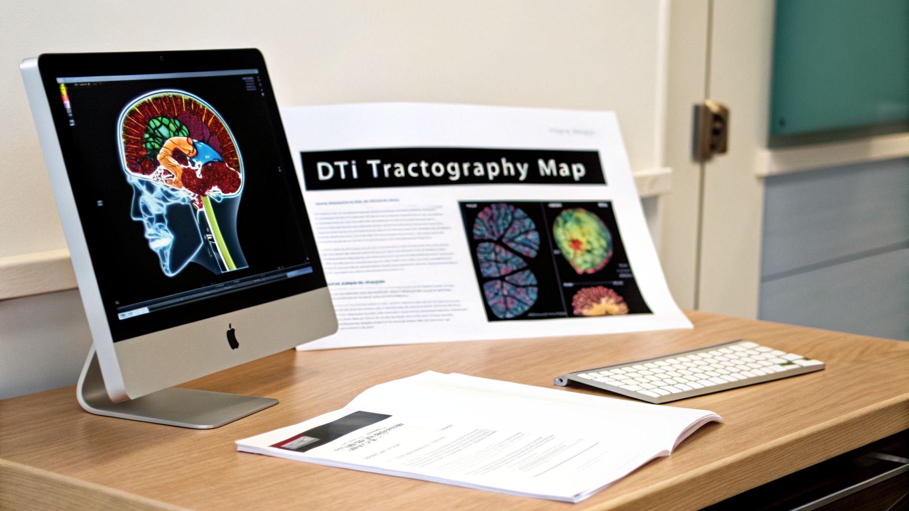
Think of the brain’s white matter as a massive, intricate highway system. A diffusion tensor imaging of the brain (DTI) scan doesn’t just give you a static map; it shows you the flow of traffic—or, in this case, the flow of water molecules along these neural highways. The result is a stunning 3D image called a tractography, which visualizes the brain's "wiring" and communication pathways.
For someone living with the aftermath of a traumatic brain injury, this is incredibly significant. Instead of just describing your symptoms, a DTI scan can show the physical disruption in your brain’s communication network. These detailed maps make an invisible injury visible, providing concrete evidence that connects the damage to your real-world struggles with memory, focus, or personality changes.
Interpreting Your DTI Results
When a neuroradiologist reviews a DTI scan, they’re looking for two main red flags we touched on earlier: Fractional Anisotropy (FA) and Mean Diffusivity (MD). By comparing your brain's metrics to established norms for a healthy brain, they can pinpoint areas of damage with remarkable accuracy.
A comprehensive DTI report will typically include:
- Color-Coded 3D Maps: These images use color to represent the direction of the brain’s pathways. Areas that look disorganized or have faded colors often signal a loss of structural integrity.
- Quantitative Data Tables: This is the raw data, showing the specific FA and MD numbers for different brain tracts. Values that fall outside the normal range are flagged as potential injury sites.
- A Narrative Report: The neuroradiologist will provide a written analysis, explaining their findings and connecting the damaged areas to the brain functions they control.
This combination of powerful visuals and hard data paints a clear picture of how the injury has impacted your brain's ability to do its job.
Understanding Key DTI Metrics After a TBI
The table below breaks down the primary measurements from a DTI scan and what they typically indicate in the context of a traumatic brain injury.
| DTI Metric | What It Measures | What It Means in a Healthy Brain | What It Means After a Brain Injury |
|---|---|---|---|
| Fractional Anisotropy (FA) | The directionality of water diffusion along white matter tracts. | High FA Value: Water is flowing in an organized, uniform direction, indicating healthy, intact nerve fibers. | Low FA Value: Water is diffusing randomly, signaling that the nerve fibers are damaged, disorganized, or destroyed. |
| Mean Diffusivity (MD) | The total amount of water diffusion in a given area, regardless of direction. | Normal MD Value: Indicates a normal density of brain tissue and cellular structure. | High MD Value: Suggests increased space between cells, often due to swelling (edema) or tissue loss (atrophy). |
Looking at these metrics together gives doctors and legal experts a scientifically-backed story of what happened inside the brain.
The Power of DTI Metrics in a Legal Case
In a legal context, these numbers become powerful pieces of evidence. For instance, a low Fractional Anisotropy (FA) value directly indicates that the axons—the brain’s communicators—have been damaged, stretched, or torn. This is the classic signature of a diffuse axonal injury (DAI).
On the other hand, a high Mean Diffusivity (MD) value often points to the after-effects of trauma, like swelling or cell death. When a neuroradiologist finds low FA and high MD in the same region, it creates a compelling, scientifically validated picture of injury.
For an accident victim, a DTI scan changes everything. It’s no longer your word against an insurance adjuster’s doubts. The DTI report provides objective, scientific proof that your brain was physically altered by the trauma.
This is especially critical in moderate to severe TBI cases, where DTI can identify and quantify diffuse axonal injury in an estimated 20-50% of patients. This gives legal teams the hard data needed to prove the existence of an "invisible" injury. You can read more about the clinical utility of DTI in traumatic brain injuries to see just how established this science has become.
Using DTI Scans in a Personal Injury Case

This is where advanced medical science becomes your strongest legal ally. For countless accident survivors, the most maddening part of their journey is being told their MRI or CT scans are "normal." All the while, you're battling debilitating headaches, memory fog, and a personality that no longer feels like your own.
This isn't just frustrating; it’s a standard tactic used by insurance companies to deny or minimize a traumatic brain injury.
Insurance adjusters love to argue that without a visible bleed or fracture, your symptoms must be psychological, pre-existing, or imagined. A diffusion tensor imaging of the brain (DTI) scan cuts through that defense. It provides the objective, scientific evidence needed to show what’s really going on, turning a disputed claim into a fact-based case for fair compensation.
Turning an Invisible Injury Into Concrete Proof
In any injury claim, you have to prove causation—that the other party’s negligence directly caused your harm. When a standard MRI comes back clean, proving this link for a brain injury becomes incredibly difficult. The defense will jump on the opportunity to claim your symptoms are all in your head.
A DTI scan closes that gap. By revealing microscopic damage to the brain’s white matter tracts, it draws an undeniable line between the physical forces of the accident and the physical changes in your brain.
- Objective Evidence: A DTI scan doesn’t rely on subjective reports. It generates hard data, like reduced FA values, that scientifically proves a physical injury exists.
- Visual Proof: Tractography maps create powerful, compelling images. We can show a judge or jury exactly where your brain’s communication network was broken.
- Expert Testimony: A skilled neuroradiologist can take the stand, use the DTI results, and explain precisely how the accident caused the specific axonal shearing seen on the scans.
This level of proof makes it nearly impossible for an insurance company to keep claiming the injury isn't real. It forces the conversation away from a debate over your symptoms and toward a discussion about documented brain damage. For a deeper look into these legal fights, you might want to read our guide on litigating traumatic brain injury cases.
In the courtroom, a DTI scan transforms your "invisible injury" into visible, undeniable proof. It validates your experience and backs up your claim with science that insurance companies can't easily dismiss.
Securing Fair Compensation for a Lifetime of Impact
Proving the injury is only the first step. A DTI scan is also crucial for showing the full, long-term impact of that injury. The results help demonstrate the severity and permanence of the damage, justifying the true value of your claim—including future medical care, lost earning capacity, and the profound pain and suffering you’ve endured.
For families staring down a lifetime of challenges after an accident, this kind of objective evidence is everything. It quantifies what the insurance company wants to ignore and supports your need for real resources.
DTI evidence strengthens claims for:
- Future Medical Care: By showing permanent structural damage, DTI proves you'll need ongoing support, like long-term rehabilitation, cognitive therapy, and other medical help.
- Lost Earning Capacity: If DTI shows damage to brain regions that control focus, executive function, or memory, it builds a powerful case that your ability to work and earn a living has been permanently compromised.
- Pain and Suffering: Nothing shows a jury the true impact of a brain injury like seeing the damage for themselves. The visual and scientific proof helps them understand why you deserve significant compensation for the loss of your quality of life.
Ultimately, a diffusion tensor imaging of the brain scan gives your legal team the leverage needed to demand a fair settlement or win at trial. It’s the scientific proof required to hold the people who harmed you fully accountable for what they took away.
Navigating the DTI Scan Process
When a doctor or attorney suggests a diffusion tensor imaging of the brain (DTI), it's normal to feel a mix of hope and apprehension. Knowing what the process involves can help calm those nerves. While a DTI scan gives us a remarkably deep look into your brain’s white matter, the experience for you is actually very simple.
The scan itself happens in a standard MRI machine. The real difference isn’t the big machine you see; it’s the highly specialized software that runs the scan and pieces the images together. From your perspective, it will feel just like a regular MRI. You’ll lie down on a sliding table, and your main job is just to hold as still as you can.
What the Scan Experience Is Like
Most people have a few questions about what the scan will feel like. Let's walk through it, so you know exactly what to expect.
- Duration: The scan usually lasts about 30-45 minutes. It’s a bit longer than a standard MRI because the machine is capturing incredibly detailed information about how water moves in your brain.
- Comfort: The scan is painless. You won't feel a thing. The machine does make loud knocking and buzzing noises, but you’ll get earplugs or headphones to block out most of the sound.
- Safety: A DTI scan is very safe. Unlike CT scans or X-rays, it does not use any ionizing radiation. The images are created using only strong magnets and radio waves.
The most critical part of the entire DTI process isn't the scan itself—it's the interpretation of the results. This isn't a task for a general radiologist. Getting accurate insights from a diffusion tensor imaging of the brain scan requires a highly specialized neuroradiologist with extensive experience in DTI analysis.
The Role of Your Legal and Medical Team
This is where having an experienced personal injury attorney makes all the difference. A lawyer who focuses on complex brain injury cases will already have a network of trusted medical experts. They know exactly who to send your scan to—a qualified neuroradiologist who can expertly analyze the data and write a thorough report.
But your attorney’s work doesn’t stop there. After getting the report, they translate the complex medical language—terms like reduced Fractional Anisotropy or increased Mean Diffusivity—into a clear and compelling story. They connect the objective data from the scan directly to the symptoms you experience every day, building a powerful argument for an insurance adjuster or jury.
This partnership between you, your legal team, and top-tier medical experts is what builds the foundation of your case. It’s how we make the invisible damage to your brain visible, validated, and impossible to ignore.
Key Questions for Your Doctor and Attorney
After an accident, you’re thrown into a world of appointments and paperwork that can feel completely overwhelming. To get clarity and take back control, you have to ask the right questions. It’s the only way to make sure you’re making informed choices and building a team that truly understands the complexities of a traumatic brain injury (TBI).
Feeling confident when you talk to your doctors and your lawyer is everything. Use these questions to steer the conversation and ensure powerful tools like diffusion tensor imaging of the brain are on the table for your case.
Speaking with Your Medical Team
You know something is wrong. The headaches, the brain fog, the mood swings—they’re real. But the standard MRI came back "normal," and you're left wondering if it's all in your head. It’s not. It’s time to push for a deeper look.
These questions can help you start that conversation with your neurologist or doctor.
- "Could my ongoing symptoms be caused by a white matter injury or a diffuse axonal injury?" This question cuts through the confusion. It shows your doctor you know that the most significant damage from an accident often happens on a microscopic level—something a regular MRI or CT scan will almost always miss. It re-frames your symptoms as evidence of a physical injury.
- "Is a diffusion tensor imaging (DTI) scan the right next step to figure out what's going on?" Asking for a DTI by name shows you’ve done your homework. It prompts your doctor to consider whether this advanced scan could provide the concrete, objective proof needed to explain your condition, especially when other tests have come up empty.
Asking direct questions like these changes your role. You’re no longer just a passive patient; you become an active partner in your own diagnosis, ensuring your medical team uses every tool available to find the real source of your struggles.
Partnering with Your Legal Counsel
The attorney you choose can make or break your case. This is one of the most critical decisions you'll face. You don't just need a personal injury lawyer; you need a legal partner with specific, hands-on experience using complex medical evidence like DTI to prove a brain injury.
Here are the questions you must ask any attorney you consider hiring:
- "What is your firm's track record using DTI scans in brain injury cases?" This isn't about whether they've handled TBI claims before. You need to know if they have actually taken DTI evidence, worked with neuroradiologists to interpret it, and successfully used it to win a case.
- "How do you work with medical experts to explain DTI data to a jury?" A seasoned TBI attorney will have a go-to network of trusted neuroradiologists. They should be able to walk you through their exact process for turning a dense medical report into a simple, compelling story that connects the microscopic damage to the accident.
- "How will a DTI scan help us fight back against the insurance company?" This question reveals their strategy. A great lawyer will immediately explain how DTI provides objective proof that dismantles the insurance adjuster's favorite arguments—like claiming your symptoms are all in your head or are from a pre-existing condition.
Your Questions About DTI Brain Scans, Answered
When you're dealing with an accident claim, the idea of another medical procedure can feel overwhelming. If a diffusion tensor imaging of the brain (DTI) scan has been mentioned, you’re bound to have practical questions.
Getting clear answers is the first step toward feeling in control. Let’s walk through the most common concerns about DTI scans in brain injury cases.
When Is the Right Time for a DTI Scan?
Many clients worry it’s "too late" to get a DTI scan, especially if the accident happened months or even years ago. That’s a common misconception. In reality, the timing is flexible, and a later scan can be incredibly powerful.
While DTI can spot damage right after an injury, its real strength often emerges in the chronic phase. It excels at revealing permanent, long-term changes to the brain’s white matter—the kind of structural damage that persists long after the initial trauma.
It can document the lasting consequences of the injury, like axonal loss that developed over time.
For a legal claim, a DTI scan performed long after the accident can be even more compelling. It proves the damage isn’t a temporary issue that simply healed. It shows a permanent condition that will continue to shape your life.
This ability to capture chronic damage is crucial for demonstrating the true scope of your losses and the need for future care.
Is DTI Evidence Allowed in Court?
Yes, DTI evidence is now widely accepted in courtrooms as the science has become more established and trusted. But its admission isn't automatic. It all comes down to how the evidence is gathered and presented.
To be admissible, the DTI results must be handled correctly. This means two things are non-negotiable:
- Your scans must be interpreted by a true expert. This isn't a job for a general radiologist. You need a board-certified neuroradiologist who has deep, specific experience with DTI.
- The findings must be explained clearly. Your expert witness has to walk a judge and jury through the science, showing exactly how your scans prove a physical brain injury.
An experienced traumatic brain injury law firm knows these standards inside and out. We partner with leading medical experts who can deliver scientifically sound and legally persuasive evidence, making DTI a game-changing tool in your case.
How Much Does a DTI Scan Cost, and Who Pays for It?
There’s no getting around it: a diffusion tensor imaging of the brain scan is expensive. To make matters worse, health insurance companies often try to deny coverage, calling the procedure "investigational." That financial hurdle can be discouraging.
In a personal injury claim, however, the cost should never be a barrier.
The expense of the DTI scan is a direct medical cost of your accident. It's a necessary diagnostic tool, and we treat it as part of the damages you are owed.
In a successful case, the at-fault party's insurance company is held responsible for this cost as part of your final settlement or verdict. Your attorney can often arrange for you to get the scan upfront, with the provider being paid out of the settlement, so you don’t have to worry about the bill while you’re trying to heal.
At Nares Law Group LLC, we understand both the science behind DTI and the legal strategy needed to use it effectively. If you are struggling with an invisible injury after an accident, we can help you get the objective proof you need. For a free, no-obligation consultation, contact us today through our website and get the justice you deserve.